Researchers at The University of Auckland's Maurice Wilkins Centre have mapped melanoma lymphoscintigraphy data from the Sydney Melanoma Unit (SMU) onto a 3D anatomically based
computer model of the skin and lymph nodes. This mapped data has enabled visualisation of the likely node fields
for melanoma metastasis.
Data was mapped by
Dr Hayley Reynolds at the Auckland Bioengineering Institute, supervised
by Dr Nicolas Smith and Assoc Prof Rod Dunbar. Lymphoscintigraphy data from the SMU is courtesy of Prof Roger Uren MD and Prof John Thompson MD.
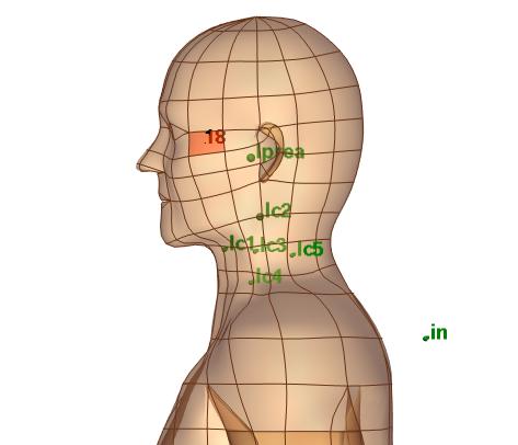
SKIN SELECTION TOOL:
Click on regions of skin on the head and neck below to display the potential sentinel lymph node fields that will receive melanoma cells if the cancer has spread
beyond the skin. The node fields displayed are scaled in size according to the drainage likelihood, and the total number of patient cases
are displayed on the selected skin regions. The table of statistics details the number of patient cases draining to each node field and the
percentage likelihood of drainage to that node field.